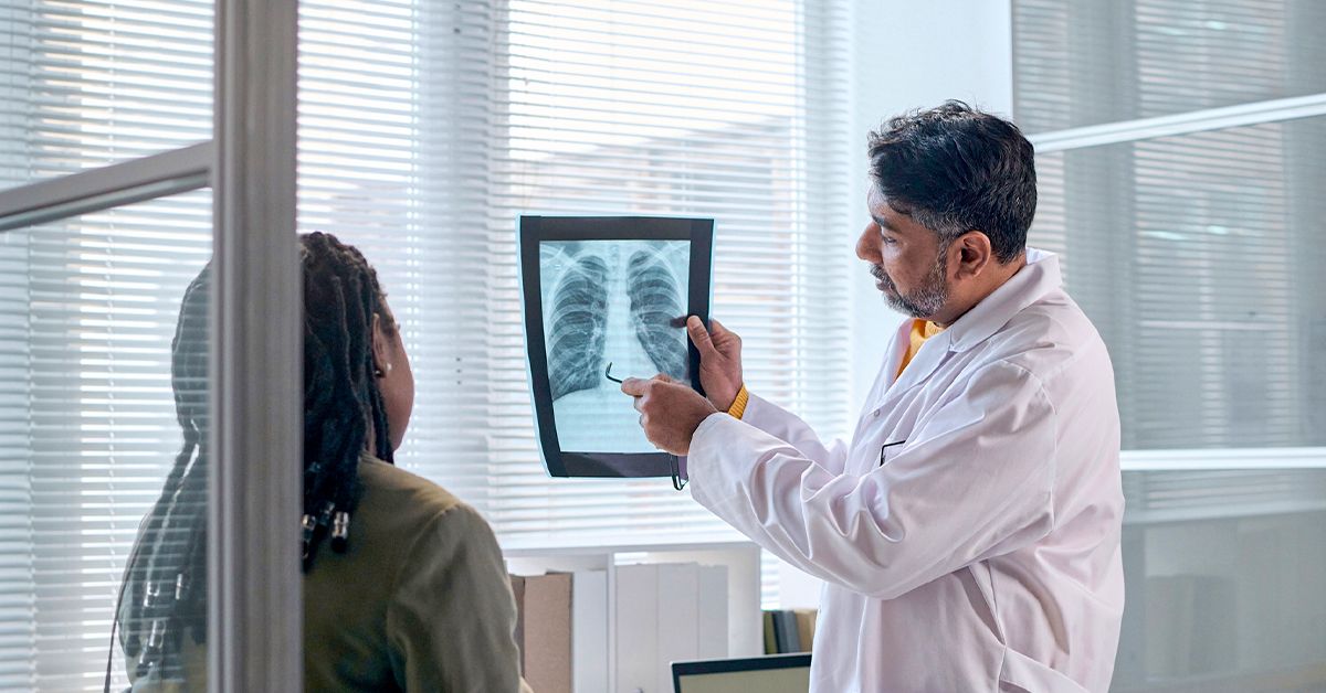
Editorial note: This article is for educational purposes only and is based on current, mainstream U.S. medical guidance. A pulmonary embolism can be life-threatening. Anyone with sudden shortness of breath, chest pain, fainting, coughing blood, or severe unexplained symptoms should seek emergency medical care immediately.
What Is a Subsegmental Pulmonary Embolism?
A subsegmental pulmonary embolism, often shortened to SSPE, is a small blood clot lodged in one of the tiny, outer branches of the pulmonary arteries. These arteries carry blood from the heart to the lungs so the blood can pick up oxygen. When a clot blocks even a small branch, it can interrupt blood flow and trigger symptoms such as chest pain, shortness of breath, or a racing heartbeat.
Think of the lung’s blood vessel system like a tree. The main pulmonary artery is the trunk, the segmental arteries are larger branches, and the subsegmental arteries are the smaller twigs. A clot in a twig may sound less dramatic than a clot in the trunk, but doctors still take it seriously. Small does not automatically mean harmless. The real question is whether the clot is isolated, whether there are more clots hiding elsewhere, and whether the person has risk factors that make another clot likely.
Subsegmental pulmonary embolism has become a more common diagnosis because modern CT pulmonary angiography can detect smaller clots than older imaging could. That is helpful, but it also creates a clinical puzzle: some patients clearly need blood thinners, while carefully selected low-risk patients may be watched closely instead of treated immediately. In other words, SSPE is not a “one-size-fits-all” diagnosis. It is more like a medical choose-your-own-adventure book, except the doctor really should be the one turning the pages.
Why Subsegmental Pulmonary Embolism Matters
Any pulmonary embolism matters because it can reduce oxygen levels, strain the right side of the heart, and signal that the body is forming dangerous clots. A subsegmental PE may be small, but it can still be connected to a deep vein thrombosis, or DVT, in the leg or pelvis. If a DVT is present, more clot material could break off and travel to the lungs.
SSPE also matters because treatment decisions involve a careful balance. Anticoagulant medications, commonly called blood thinners, can prevent clots from growing and help stop new ones from forming. However, they can also increase bleeding risk. That is why clinicians look at the whole picture: symptoms, imaging results, leg ultrasound findings, cancer status, pregnancy, recent surgery, previous clot history, oxygen levels, heart strain, bleeding risk, and the reliability of follow-up care.
Common Causes of Subsegmental Pulmonary Embolism
Most pulmonary emboli begin as clots in deep veins, especially in the legs. When part of a clot breaks loose, it can travel through the bloodstream, pass through the right side of the heart, and lodge in the lung arteries. In SSPE, the clot ends up in a smaller, more distant artery.
Deep Vein Thrombosis
DVT is one of the most important causes of pulmonary embolism. A clot may form in the calf, thigh, pelvis, or sometimes the arm. Symptoms of DVT can include swelling, tenderness, warmth, redness, or pain in one limb. However, DVT can also be sneaky and cause few or no symptoms. That is why doctors often order compression ultrasound of the legs when SSPE is found. If a proximal DVT is present, anticoagulation is usually recommended.
Immobility and Long Periods of Sitting
Blood likes to move. When it slows down, it becomes more likely to clot. Long flights, long car rides, bed rest, hospitalization, or sitting for extended periods can raise clot risk. This does not mean everyone needs to panic after a road trip, but it does mean movement matters. Calf muscles are not just decorative; they help pump blood back toward the heart.
Surgery, Injury, and Hospitalization
Major surgery, especially hip, knee, abdominal, pelvic, or cancer-related surgery, can increase the risk of blood clots. Trauma and serious injury can do the same. Hospitalized patients may have several risk factors at once: reduced mobility, inflammation, IV lines, infection, and underlying illness.
Cancer and Cancer Treatment
Cancer can make the blood more prone to clotting. Some chemotherapy, surgery, central venous catheters, and reduced mobility add even more risk. In a person with active cancer, even a small subsegmental PE is usually taken very seriously because the risk of recurrence may be higher.
Pregnancy, Postpartum Period, and Estrogen Exposure
Pregnancy and the weeks after delivery increase clot risk because of normal changes in blood clotting, pressure on pelvic veins, and reduced mobility for some patients. Estrogen-containing birth control or hormone therapy can also raise the chance of venous thromboembolism in certain people, especially when combined with smoking, inherited clotting disorders, or previous clots.
Inherited or Acquired Clotting Disorders
Some people inherit clotting tendencies, such as factor V Leiden or prothrombin gene mutation. Others develop acquired conditions, such as antiphospholipid syndrome. Testing is not needed for every person with SSPE, but it may be considered when clots occur at a young age, happen repeatedly, appear without an obvious trigger, or are associated with a strong family history.
Symptoms of Subsegmental Pulmonary Embolism
Subsegmental PE symptoms can range from obvious to barely noticeable. Some people feel suddenly short of breath. Others have sharp chest pain that worsens with a deep breath, a symptom called pleuritic chest pain. Some feel unusually anxious, lightheaded, sweaty, or aware of a fast heartbeat. A cough may occur, and occasionally the cough brings up blood.
Common symptoms may include:
- Sudden or unexplained shortness of breath
- Chest pain, especially pain that worsens with deep breathing or coughing
- Rapid or irregular heartbeat
- Lightheadedness, dizziness, or fainting
- Cough, sometimes with blood-streaked mucus
- Leg swelling, calf pain, warmth, or redness if DVT is present
- Unusual fatigue or reduced exercise tolerance
Not everyone with SSPE has dramatic symptoms. Some cases are discovered while doctors are looking for something else on a CT scan. Still, symptoms should never be dismissed just because the clot is “small.” If the lungs and heart are involved, the body deserves attention, not a pep talk and a shrug.
When to Seek Emergency Care
Emergency care is needed for sudden shortness of breath, chest pain, fainting, coughing blood, blue or gray lips, severe weakness, confusion, or very low blood pressure. These symptoms can signal a serious pulmonary embolism, heart attack, severe infection, collapsed lung, or another urgent condition. It is not possible to reliably sort these out at home with a search engine, a smartwatch, and wishful thinking.
People with known clot risk factors should be especially cautious. That includes those with recent surgery, recent hospitalization, active cancer, pregnancy or recent delivery, previous DVT or PE, major injury, long travel, or a known clotting disorder.
How Doctors Diagnose Subsegmental Pulmonary Embolism
Diagnosis usually starts with a medical history, physical exam, oxygen level, heart rate, and assessment of risk. Clinicians may use structured tools such as Wells score, Geneva score, pulmonary embolism rule-out criteria, D-dimer testing, and imaging. The goal is to avoid missing a dangerous clot while also avoiding unnecessary testing and treatment.
D-Dimer Blood Test
D-dimer is a blood test that can rise when the body is breaking down clots. A normal D-dimer can help rule out PE in low-risk patients. However, D-dimer can be elevated for many reasons, including infection, pregnancy, inflammation, recent surgery, trauma, cancer, and older age. In other words, D-dimer is sensitive, but it is not a mind reader.
CT Pulmonary Angiography
CT pulmonary angiography, or CTPA, is the most common imaging test used to diagnose pulmonary embolism. It uses contrast dye to show blood flow in the pulmonary arteries. CTPA is very good at finding clots, including small subsegmental clots, but interpretation can sometimes be challenging. Tiny abnormalities may be real clots, artifacts, or findings of uncertain significance, which is one reason expert review matters.
Ventilation-Perfusion Scan
A ventilation-perfusion scan, also called a V/Q scan, compares airflow and blood flow in the lungs. It may be used when CTPA is not ideal, such as in people with severe kidney disease, contrast allergy, or certain pregnancy-related situations. A mismatch between ventilation and blood flow can suggest pulmonary embolism.
Leg Ultrasound
Because many pulmonary emboli start as DVT, ultrasound of the legs is often important. If ultrasound finds a proximal DVT, that strongly supports treatment with anticoagulation, even if the lung clot is small.
Treatment Options for Subsegmental Pulmonary Embolism
Treatment for SSPE depends on risk. Some patients need anticoagulation. Some may need hospital monitoring. A smaller group of carefully selected patients may be managed with structured surveillance instead of immediate blood thinners.
Anticoagulant Medication
Anticoagulants are the main treatment for most pulmonary emboli. They do not “melt” a clot instantly. Instead, they prevent the clot from growing and reduce the chance of new clots while the body gradually breaks down the existing clot. Common options include direct oral anticoagulants, low molecular weight heparin, unfractionated heparin, fondaparinux, and warfarin. For many eligible adults with acute PE, direct oral anticoagulants are commonly preferred because they are effective and convenient, but they are not right for everyone.
Patients with kidney disease, liver disease, pregnancy, antiphospholipid syndrome, high bleeding risk, medication interactions, extreme body weight, or active cancer may need a different plan. This is why copying someone else’s blood thinner regimen from an online forum is a terrible hobby.
Structured Surveillance Without Immediate Anticoagulation
In selected low-risk patients with isolated subsegmental PE, no proximal DVT, good cardiopulmonary reserve, low risk of recurrence, low symptom burden, and reliable follow-up, doctors may consider surveillance instead of anticoagulation. Surveillance usually includes close clinical follow-up and repeat leg ultrasound to make sure a DVT does not appear or progress.
This approach is not casual “wait and see.” It is organized, documented, and monitored. It is generally not favored when a patient has active cancer, pregnancy, multiple subsegmental emboli, poor heart or lung reserve, marked symptoms, previous unprovoked VTE, or unreliable access to follow-up.
Hospitalization vs. Home Treatment
Some low-risk PE patients may be treated at home with careful instructions and follow-up. Others need hospital care, especially if they have low oxygen levels, low blood pressure, heart strain, severe symptoms, high bleeding risk, complex medical conditions, or limited support at home. The location of care depends less on the word “subsegmental” and more on the patient’s overall stability.
Thrombolysis, Catheter Procedures, and Surgery
Clot-busting drugs, catheter-directed therapy, thrombectomy, or surgical embolectomy are generally reserved for severe pulmonary embolism with shock, hemodynamic instability, or serious deterioration. These treatments are not typical for an isolated low-risk subsegmental PE, but they may be considered in life-threatening PE situations.
How Long Does Treatment Last?
When anticoagulation is used, treatment often lasts at least three months, but the exact duration depends on why the clot happened and the risk of recurrence. A clot triggered by a temporary risk factor, such as surgery, may require a different duration than an unprovoked clot or a clot linked to ongoing cancer. Some patients need extended therapy, while others stop after a defined course.
Follow-up is important. Patients may need monitoring for bleeding, medication interactions, kidney function, symptom improvement, and signs of recurrence. Some people also need evaluation for chronic thromboembolic disease if shortness of breath or exercise limitation continues after treatment.
Possible Complications
Many people recover well from pulmonary embolism, especially when it is recognized and managed properly. Still, complications can occur. A clot may recur, bleeding may happen during anticoagulant therapy, or persistent symptoms may develop. Rarely, unresolved clots can contribute to chronic thromboembolic pulmonary hypertension, a condition in which pressure in the lung arteries stays high and strains the heart.
Warning signs during recovery include worsening shortness of breath, new chest pain, fainting, new leg swelling, black or bloody stools, severe headaches, unusual bruising, or bleeding that does not stop. Anyone taking anticoagulants should know what bleeding symptoms to watch for and when to call a clinician.
Prevention: How to Lower the Risk of PE
Preventing subsegmental pulmonary embolism starts with reducing the risk of venous blood clots. Prevention is especially important after surgery, during hospitalization, during long travel, and for people with previous VTE.
Move Early and Often
During long trips, walking breaks, calf exercises, hydration, and avoiding long stretches of stillness can help. After surgery or illness, follow the medical team’s instructions about safe movement. The goal is not to run a marathon in the hallway; it is to keep blood moving.
Use Preventive Medication When Prescribed
Some patients receive preventive anticoagulants after surgery, during hospitalization, or during high-risk periods. These medications should be taken exactly as directed. Skipping doses can reduce protection, while doubling up can raise bleeding risk.
Know Personal Risk Factors
People with previous DVT or PE, active cancer, clotting disorders, family history, obesity, smoking, inflammatory disease, or estrogen therapy should discuss clot prevention with a healthcare professional. Risk is personal, and prevention works best when it is tailored.
Living With and Recovering From Subsegmental Pulmonary Embolism
Recovery from SSPE can feel confusing because the diagnosis may sound small, but the emotional impact can be large. Many patients hear “blood clot in the lung” and immediately picture the worst. Others hear “subsegmental” and wonder whether it was serious at all. The truth usually sits in the middle: it is a real diagnosis that deserves respect, but many people do very well with the right care plan.
A practical recovery plan often includes taking medication as prescribed, attending follow-up visits, asking whether repeat imaging or ultrasound is needed, watching for bleeding symptoms, and gradually returning to normal activity. Some people feel better quickly. Others need weeks or months before stamina fully returns. Fatigue after PE is not unusual, and it can be frustrating for people who are used to bouncing back fast.
Experiences Related to Subsegmental Pulmonary Embolism: What Patients Often Notice
Many people who go through a subsegmental pulmonary embolism describe the experience as “strange” rather than dramatic. Unlike a broken bone, where the problem is visible and obvious, SSPE can feel invisible. One person may say, “I just felt winded walking up the stairs.” Another may say, “It felt like a sharp pinch when I breathed in.” Someone else may have no major symptoms and learn about the clot after a CT scan for chest discomfort, pneumonia-like symptoms, or follow-up after another condition. That uncertainty can be emotionally exhausting.
A common experience is the mismatch between how small the clot sounds and how serious the diagnosis feels. Patients may leave the emergency department or clinic with a prescription for anticoagulants and a head full of questions: Can I exercise? Can I travel? What if I cut myself shaving? Is every chest twinge another clot? Should I sleep sitting up? The medical answer is usually individualized, but the emotional answer is universal: it is normal to feel cautious after a lung clot.
People treated with blood thinners often need time to adjust. Daily routines may change. They may need to avoid certain over-the-counter medications unless approved by a clinician, report unusual bleeding, check for drug interactions, and tell dentists or other healthcare professionals about anticoagulant use. For patients on warfarin, regular blood testing and dietary consistency may be part of life. For patients on direct oral anticoagulants, routine lab monitoring may be less demanding, but taking doses consistently remains essential.
Another frequent experience is learning to interpret symptoms without becoming consumed by them. Mild soreness from exercise, anxiety-related chest tightness, acid reflux, and muscle strain can all mimic scary symptoms. At the same time, new or worsening shortness of breath, fainting, coughing blood, or significant chest pain should not be ignored. Many patients find it helpful to ask their clinician for a clear “red flag” list before leaving care. A written plan can calm the mind more effectively than trying to remember instructions while wearing a hospital bracelet and pretending not to be nervous.
Family and friends may also misunderstand SSPE. Because the person may look normal, others might assume everything is fine immediately. But recovery can include fatigue, anxiety, medication concerns, and follow-up appointments. A helpful support person does not need to become a clot expert overnight. Listening, offering rides, helping organize medication schedules, and taking symptoms seriously can make a big difference.
Work and activity are another adjustment. Some people return quickly, especially if symptoms are mild and the job is not physically demanding. Others need modified duties or a gradual return. Exercise is usually encouraged once a clinician says it is safe, but the pace should be realistic. The goal is steady recovery, not proving anything to the treadmill. The treadmill, frankly, has no feelings.
Finally, many patients become more aware of prevention after SSPE. They stand up during long travel, hydrate better, ask about clot prevention before surgery, and recognize DVT symptoms sooner. That awareness is not paranoia; it is informed self-care. A subsegmental pulmonary embolism can be frightening, but it can also become the moment a person learns how to protect their circulation, communicate better with clinicians, and take warning signs seriously without letting fear run the entire show.
Conclusion
Subsegmental pulmonary embolism is a small clot in a small lung artery, but its management requires big-picture thinking. The right treatment depends on whether the clot is truly isolated, whether DVT is present, how severe the symptoms are, and whether the patient has risk factors for recurrence or bleeding. Some patients need anticoagulants, while carefully selected low-risk patients may be candidates for structured surveillance. Either way, SSPE should be evaluated by qualified healthcare professionals, not guessed at from symptoms alone.
The best takeaway is simple: know the warning signs, understand the risk factors, follow the treatment plan, and keep follow-up appointments. A small clot can still carry important information about your health. Listening to that message early can help prevent bigger problems later.