Cancer detection has never been a casual game of “spot the difference.” For decades, doctors have relied on imaging scans, biopsies, pathology reports, lab tests, and a healthy amount of professional detective work to find tumors, identify their behavior, and decide what to do next. The problem is that cancer does not always politely wave from the front row. Sometimes it hides in dense tissue, sheds tiny molecular clues into the bloodstream, or grows in places where symptoms arrive fashionably latefar too late, unfortunately.
That is why real-time cancer detection is becoming one of the most exciting frontiers in modern oncology. Instead of waiting days or weeks for answers, the future points toward faster signals: artificial intelligence that flags suspicious images, liquid biopsy tests that track circulating tumor DNA, fluorescence-guided surgery that helps surgeons see tumor margins, and digital pathology tools that scan tissue slides with tireless attention. In simple terms, oncology is moving from “let’s wait for the report” toward “let’s understand what is happening right now.” That shift could change cancer screening, diagnosis, treatment planning, surgery, follow-up care, and even how patients experience the entire journey.
What Does Real-time Cancer Detection Mean?
Real-time cancer detection does not mean one magical scanner that instantly declares, “Aha! Cancer!” Medicine is impressive, but it has not turned into a sci-fi toaster yet. Instead, the phrase refers to technologies that shorten the time between cancer signal, clinical interpretation, and medical action. These tools may help identify suspicious tissue during surgery, detect tumor DNA in blood, highlight abnormalities on scans, or monitor whether treatment is working before a tumor visibly changes size.
At its best, real-time detection combines speed with accuracy. The goal is not merely to get answers faster; it is to get better answers at the moment when they matter most. A surgeon deciding whether a margin is clean, an oncologist wondering whether a therapy is failing, or a radiologist reviewing hundreds of scans can all benefit from immediate, reliable support. In oncology, timing is not just convenient. It can be the difference between a smaller procedure and a bigger one, a targeted therapy and a guessing game, or early intervention and delayed action.
Why Early and Faster Detection Matters So Much
The American cancer landscape remains enormous. Millions of people in the United States are diagnosed with cancer every year, and hundreds of thousands die from it. The encouraging news is that survival has improved for many cancers because of better screening, lower smoking rates, targeted therapies, immunotherapy, and improved supportive care. The less cheerful newsbecause cancer apparently did not get the memo about being less complicatedis that some cancers are still commonly found late, when treatment becomes harder and outcomes worsen.
Traditional screening already saves lives. Mammography, colonoscopy, stool-based colorectal tests, Pap tests, HPV testing, and low-dose CT scans for eligible people have reshaped prevention and early detection. But traditional screening has limits. Some cancers lack widely recommended screening tests. Some tumors grow between scheduled exams. Some patients face access barriers, while others avoid screening because it feels confusing, expensive, uncomfortable, or simply easy to postpone until “next month,” which somehow becomes next decade.
Real-time cancer detection aims to close some of those gaps. Faster detection could help doctors find cancers earlier, personalize treatment sooner, monitor recurrence more closely, and reduce unnecessary procedures. However, speed must be paired with evidence. A fast wrong answer is not innovation; it is just a mistake wearing sneakers.
Artificial Intelligence: The New Second Set of Eyes
Artificial intelligence is quickly becoming one of the most discussed tools in cancer detection. In radiology, AI can help analyze mammograms, CT scans, MRIs, and other images by highlighting suspicious patterns. In pathology, AI can scan digitized tissue slides and assist with tumor grading, cell counting, and pattern recognition. In oncology research, AI can connect imaging, genomics, lab results, and clinical history to help predict risk or treatment response.
Think of AI as a very fast assistant with excellent eyesight and no need for coffee breaks. That does not mean it replaces physicians. A good oncology AI system should support trained professionals, not wander into the clinic wearing a tiny digital lab coat and making solo decisions. The strongest use cases are those where AI reduces workload, improves consistency, catches subtle findings, and helps clinicians prioritize cases that need urgent attention.
AI in Medical Imaging
Medical imaging is one of the clearest homes for real-time cancer detection. Radiologists review enormous volumes of images, and small lesions can be difficult to spot, especially when they are early, faint, or hidden among normal anatomy. AI tools can help flag lung nodules, suspicious breast findings, prostate lesions, brain tumors, or liver abnormalities. These systems may also compare current and previous scans, identify growth patterns, and support risk scoring.
For lung cancer, the potential is especially important. Low-dose CT screening can reduce mortality in high-risk groups, but screening programs require accurate reading, follow-up management, and access. AI could help identify nodules earlier and standardize reporting, particularly in busy hospitals or underserved regions. The technology is not perfect, but it can act like a smart spotlight: it does not perform the play, but it helps everyone see the stage.
AI in Digital Pathology
Pathology has traditionally depended on glass slides and microscopes. Digital pathology changes that by converting slides into high-resolution images that can be analyzed, shared, archived, and reviewed with computational tools. AI may help identify cancer cells, measure biomarkers, detect lymph node metastases, estimate tumor grade, or quantify features that are difficult for humans to count consistently.
This matters because pathology is central to diagnosis. A scan may suggest cancer, but tissue often confirms it. Faster pathology workflows can reduce waiting time and help oncologists move from suspicion to treatment planning sooner. For patients, that waiting period can feel like time is moving through cold peanut butter. Anything that responsibly shortens uncertainty is not just technically useful; it is emotionally meaningful.
Liquid Biopsy: Reading Cancer Clues in Blood
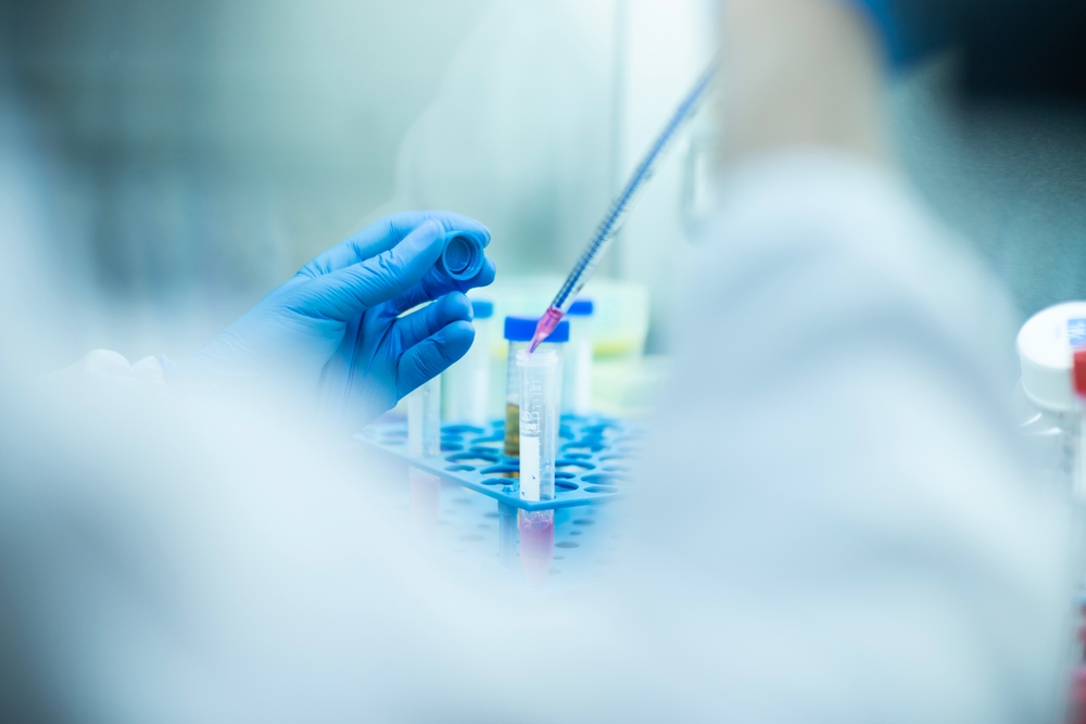
Liquid biopsy is another major pillar of real-time oncology. Instead of removing tissue from a tumor, a liquid biopsy analyzes blood or other body fluids for cancer-related signals. These may include circulating tumor DNA, circulating tumor cells, RNA, proteins, methylation patterns, or other biomarkers. The most famous character in this cast is ctDNA, tiny fragments of tumor genetic material that can enter the bloodstream.
Liquid biopsy is already useful in certain treatment settings, especially for advanced cancers where doctors need to identify mutations, choose targeted therapy, detect resistance, or monitor disease over time. For example, in lung cancer, blood-based genomic testing may help identify actionable mutations when tissue is difficult to obtain. In colorectal, breast, lung, and other cancers, ctDNA monitoring is being studied for minimal residual disease, recurrence risk, and early treatment response.
The dream is simple: a blood draw that tells doctors what a tumor is doing before scans show obvious changes. The reality is more complicated. Early tumors may shed very little DNA. False positives and false negatives can occur. A positive blood signal may not always reveal exactly where cancer is located. And multi-cancer early detection tests are still being evaluated for how they should be used, who should receive them, and whether they improve long-term outcomes.
Multi-cancer Early Detection Tests: Promise with Guardrails
Multi-cancer early detection tests, often called MCED tests, are designed to look for signals from multiple cancer types in a single blood sample. That sounds wonderfully efficientlike ordering one test and getting the medical equivalent of a full buffet. But clinical usefulness depends on more than technological coolness. The key questions are: Does the test find dangerous cancers early enough to change outcomes? How often is it wrong? What happens after a positive result? Can patients access follow-up care? Will it reduce deaths, or mostly create anxiety and extra procedures?
Current MCED research is exciting, especially for cancers that lack routine screening. Studies have shown that cancer-related DNA can sometimes be detected long before clinical diagnosis. That raises the possibility of earlier intervention, especially for pancreatic, ovarian, liver, and other cancers that are often diagnosed late. Still, these tests should not be treated as replacements for recommended screening. A blood test should not become an excuse to skip colonoscopy, mammography, cervical screening, or lung screening when those are appropriate. The future is likely additive, not either-or.
Real-time Detection During Surgery
One of the most practical uses of real-time cancer detection happens in the operating room. Surgeons often need to remove a tumor while preserving as much healthy tissue as possible. The challenge is knowing where the cancer ends and normal tissue begins. Sometimes the boundary is obvious. Sometimes it is about as clear as a foggy windshield after a drive-through car wash.
Fluorescence-guided surgery and other intraoperative imaging techniques aim to make tumor tissue more visible during the procedure. A tumor-targeting dye or imaging agent may help highlight cancer cells under special cameras. Surgeons can then assess margins, identify small deposits, and potentially reduce the need for repeat operations. This is especially relevant in cancers where complete removal improves outcomes but overly aggressive removal can damage function or quality of life.
Real-time surgical guidance is still evolving. The success of these methods depends on the imaging agent, tumor biology, camera system, workflow, and surgeon experience. But the direction is clear: future operating rooms may become more like intelligent navigation systems, helping surgeons see molecular information instead of relying only on anatomy and touch.
Monitoring Treatment Response in Real Time
Real-time cancer detection is not only about finding cancer at the beginning. It also matters after treatment starts. Historically, oncologists often waited for follow-up scans to determine whether chemotherapy, immunotherapy, radiation, or targeted therapy was working. But tumors can be sneaky. Some shrink slowly. Some appear stable before progressing. Some develop resistance at the molecular level before imaging reveals the problem.
ctDNA monitoring may help reveal treatment response earlier. If tumor DNA levels fall after therapy, that may suggest the cancer burden is decreasing. If ctDNA rises, it may signal resistance, recurrence, or minimal residual disease. Researchers are studying how to use this information to escalate treatment, de-escalate therapy, or switch drugs earlier. In the future, oncology may become less like driving by looking in the rearview mirror and more like using a dashboard with live alerts.
The Future Oncology Workflow: Faster, Smarter, More Personal
The future of oncology will likely combine multiple data streams. A patient may have imaging analyzed by AI, a biopsy evaluated with digital pathology, genomic sequencing to identify mutations, liquid biopsy to monitor tumor evolution, and wearable data to track symptoms or functional status. Instead of one snapshot, doctors may see a moving picture of cancer behavior.
This is where precision oncology becomes more precise. Cancer is not one disease; it is a large, badly behaved family of diseases with different drivers, weaknesses, and escape routes. Real-time detection can help doctors understand not only where cancer is, but what it is doing. That may support more individualized decisions: who needs aggressive treatment, who may avoid unnecessary therapy, who is at high risk of recurrence, and who should enter a clinical trial.
Challenges: Accuracy, Equity, Privacy, and Trust
Every powerful technology brings challenges. In cancer detection, the stakes are high because errors can lead to delayed care, unnecessary anxiety, invasive procedures, or inappropriate treatment. AI models must be validated in diverse populations. Liquid biopsy tests must prove clinical value, not just technical brilliance. Digital tools must integrate smoothly into real hospital workflows. Data privacy must be protected. Patients need clear explanations that do not sound like they were written by a robot trapped in a filing cabinet.
Equity is one of the biggest issues. Real-time detection should not become a luxury upgrade available only to patients at wealthy medical centers. Rural hospitals, community clinics, safety-net systems, and underserved populations must be included in research, implementation, and reimbursement planning. If innovation widens disparities, it has failed part of its mission. The best future is not just high-tech; it is widely reachable.
What Patients Should Understand Today
Patients do not need to memorize every biomarker acronym to benefit from progress. A few practical points matter. First, recommended cancer screening still matters. Second, new tests should be discussed with a qualified clinician, especially if they are not yet approved for broad screening use. Third, AI-assisted results still require medical interpretation. Fourth, a negative test does not always mean zero risk, and a positive test does not always mean cancer is definitely present.
The best patient question may be simple: “How will this test change what we do next?” If the answer is clear, evidence-based, and connected to a reasonable follow-up plan, the test may be useful. If the answer is a shrug wearing a white coat, caution is wise.
Specific Examples Shaping the Field
Several real-world examples show where oncology is heading. In breast imaging, AI-assisted mammography is being tested as a way to improve detection and reduce radiologist workload. In lung cancer, AI tools can help identify suspicious nodules on CT scans and support earlier follow-up. In pancreatic cancer research, AI models are being studied to find subtle signals that may appear before diagnosis. In advanced cancer care, liquid biopsy can identify mutations when tissue biopsy is difficult or insufficient. In surgery, fluorescence imaging may help surgeons see tumor boundaries more clearly.
These examples are not isolated gadgets. They are pieces of a larger transformation. Oncology is becoming more continuous, more data-rich, and more adaptive. The old model often asked, “What did the cancer look like at diagnosis?” The new model increasingly asks, “What is the cancer doing now, and what should we do before it gets ahead of us?”
The Human Side of Faster Detection
Technology gets the headlines, but patients live the waiting. Waiting for a scan result. Waiting for biopsy confirmation. Waiting to learn if treatment worked. Waiting to know whether “something suspicious” is harmless or life-changing. Real-time cancer detection has emotional value because it can reduce uncertainty. Not always, not perfectly, but meaningfully.
At the same time, faster information can create faster anxiety. A blood test that finds a vague cancer signal may start a stressful diagnostic journey. An AI flag on a scan may lead to additional imaging. A sensitive test may find abnormalities that never would have caused harm. That is why the future of oncology needs not only better machines, but better communication. Patients deserve plain-language explanations, shared decision-making, and care teams that understand both the biology and the human being sitting in the exam room.
Experience-Based Perspectives: What Real-time Detection Feels Like in Practice
In practical oncology settings, the most noticeable change is not always dramatic. It often begins with workflow. A radiology team receives AI-prioritized scans, and the most suspicious cases move faster through the queue. A pathologist reviews a digitized slide with computational assistance, spotting areas that deserve closer inspection. An oncologist checks molecular test results before choosing therapy instead of relying only on tumor location. These steps may sound modest, but medicine often improves through many small time savings that add up to better decisions.
Consider a patient with a suspicious lung nodule. In a traditional pathway, the person may move from scan to specialist referral to biopsy to molecular testing to treatment planning. Each step can take time. With better real-time systems, imaging software may help classify the nodule, scheduling systems may prioritize follow-up, liquid biopsy may provide molecular clues if tissue is hard to obtain, and the oncology team may discuss targeted options sooner. The patient still needs expert care, but the process becomes less like wandering through a maze while holding a paper map upside down.
Another experience comes from treatment monitoring. Patients receiving targeted therapy or immunotherapy often want to know whether the treatment is working. Scans are useful, but they are periodic. Symptoms can be misleading. Blood-based monitoring may one day give earlier clues about response or resistance. For a patient, that could mean fewer weeks spent wondering. For a doctor, it could mean adjusting therapy before the disease visibly progresses. For the healthcare system, it could mean avoiding ineffective treatment sooner, which is better for both outcomes and costs.
Surgery offers a different kind of experience. Imagine removing a tumor and being able to see suspicious tissue light up in real time. That does not make surgery easysurgeons are not playing a video game with cheat codesbut it may improve confidence around margins and reduce the chance that cancer cells are left behind. Patients may benefit from fewer repeat operations, better tissue preservation, and more precise procedures. In cancers where millimeters matter, better visualization is a big deal.
There is also an experience lesson from patient communication. Real-time detection tools can produce complex results, and complex results can confuse people quickly. A report that says “variant detected,” “indeterminate signal,” or “AI suspicion score” may be clinically useful, but it can sound terrifying without context. The future of oncology must include better result design: clear language, risk explanation, next steps, and support. A brilliant test with poor communication is like a smoke alarm that only speaks in riddles.
The most realistic experience-based takeaway is this: real-time cancer detection will not remove uncertainty from oncology, but it can shrink it. It can help doctors act earlier, patients understand sooner, and care teams personalize treatment more intelligently. The future will not be one blood test, one algorithm, or one glowing surgical dye. It will be a connected system where evidence-based tools work together, humans remain in charge, and cancer has fewer places to hide.
Conclusion: The Future Is Real-time, But It Must Be Responsible
Real-time cancer detection represents a major shift in oncology. AI, liquid biopsy, digital pathology, fluorescence-guided surgery, and continuous monitoring are moving cancer care toward earlier insight and faster action. These technologies could help find cancer sooner, guide treatment more precisely, detect recurrence earlier, and reduce unnecessary procedures. That is the bright sideand it is genuinely bright.
But the future must be built carefully. New tools need strong evidence, transparent regulation, equitable access, privacy protection, and thoughtful clinical use. Faster detection is valuable only when it leads to better decisions and better outcomes. The goal is not to turn every patient into a walking data stream. The goal is to give doctors and patients clearer signals at the moments when those signals can change lives.
Note: This article is for educational publishing purposes only and should not replace professional medical advice, diagnosis, screening recommendations, or treatment decisions from a licensed healthcare professional.