Note: This guide is for educational reading only. An ultrasound image should always be interpreted by a licensed radiologist, sonographer, obstetrician, or other qualified healthcare professional. In other words, enjoy learning the mapbut let the trained pilot land the plane.
Introduction: Why Ultrasound Pictures Look Like Weather Reports From the Moon
At first glance, an ultrasound picture can look like a mysterious black-and-white snowstorm. There are shadows, bright lines, blurry shapes, labels, numbers, and sometimes a tiny hand waving as if the baby already knows everyone is confused. But once you understand the basics, a sonogram becomes much less intimidating.
Ultrasound imaging, also called sonography, uses high-frequency sound waves to create real-time images of structures inside the body. Unlike X-rays or CT scans, ultrasound does not use ionizing radiation, which is one reason it is commonly used during pregnancy, abdominal exams, heart evaluations, pelvic imaging, breast imaging, vascular studies, and many other medical checks.
This article explains how to read an ultrasound picture in 9 steps, using simple American English, practical examples, and enough medical context to help you understand what you are looking at without pretending you suddenly graduated from radiology school during lunch.
How to Read an Ultrasound Picture: 9 Steps
Step 1: Start With the Patient and Image Labels
Before studying the image itself, look at the text around the edges. Ultrasound pictures usually include basic information such as the patient’s name or ID, date of the exam, facility name, exam type, probe setting, depth, and sometimes the body part being scanned.
For example, a pregnancy ultrasound may show labels such as “OB,” “GA,” “CRL,” “BPD,” or “FHR.” An abdominal ultrasound may include terms like “liver,” “gallbladder,” “kidney,” or “aorta.” These labels help you understand what the image is supposed to show. Without them, reading a sonogram is like opening a random page in a cookbook and trying to guess whether you are making soup or a birthday cake.
Check the date, too. A fetal ultrasound from 8 weeks of pregnancy will look completely different from a 20-week anatomy scan. A follow-up kidney ultrasound may be compared with an earlier exam to see whether a finding has changed.
Step 2: Understand the Black, White, and Gray Areas
Ultrasound images are built from echoes. The machine sends sound waves into the body, and tissues reflect those waves back differently. The result is a grayscale image.
Black areas usually represent fluid because fluid allows sound waves to pass through easily. Examples include amniotic fluid, urine in the bladder, simple cysts, or fluid-filled spaces. On a pregnancy ultrasound, the dark area around the fetus is typically amniotic fluid.
White or bright areas often represent dense structures that reflect more sound, such as bone, calcification, or strong tissue boundaries. In a fetal ultrasound, bones may appear bright. In a gallbladder ultrasound, gallstones may appear bright and may cast a shadow behind them.
Gray areas represent soft tissues, organs, muscles, and other structures with mixed echo patterns. The liver, uterus, thyroid, kidneys, and muscles all have their own texture. Radiologists often describe these appearances with terms such as “echogenic,” “hypoechoic,” “anechoic,” or “heterogeneous.”
Step 3: Learn the Basic Ultrasound Vocabulary
Medical imaging loves fancy words. Fortunately, some of the most common ultrasound terms are easier than they sound.
Anechoic means the area appears black because it produces little or no echo. Simple fluid is often anechoic.
Hypoechoic means darker than nearby tissue. Some cysts, nodules, or inflamed areas may appear hypoechoic.
Hyperechoic means brighter than nearby tissue. Fat, bone, calcification, scar tissue, or stones may appear hyperechoic.
Isoechoic means similar brightness to surrounding tissue. This can make some findings harder to see.
Acoustic shadowing means a dark shadow appears behind a bright object because sound waves cannot pass through well. Stones and bones commonly cause shadowing.
Posterior enhancement means the area behind a fluid-filled structure looks brighter because sound travels easily through fluid. This can help identify simple cysts.
Knowing these words helps you read both the ultrasound picture and the written report. It also makes you sound extremely prepared at appointments, which is always a nice bonus.
Step 4: Identify the Orientation of the Image
One of the trickiest parts of reading an ultrasound picture is orientation. The image does not always match the way you naturally imagine the body. Depending on the probe position, the top of the screen may represent the skin surface, and deeper structures appear lower on the image.
Many ultrasound images include a small marker or dot on one side. This corresponds to the marker on the ultrasound probe. Sonographers use standard scanning positions so that doctors can interpret the images consistently.
In pregnancy imaging, the baby may appear upside down, sideways, curled up, or doing what looks like advanced yoga. That does not necessarily mean anything is wrong. It simply reflects how the probe captured the image at that moment.
For abdominal ultrasound, the same organ may be scanned in different planes: longitudinal, transverse, or oblique. A kidney, for example, may look bean-shaped in one view and rounder in another. This is normal because ultrasound creates slices, not a single whole-body photograph.
Step 5: Look for the Main Structure First
Do not try to interpret every pixel at once. Start by identifying the main structure the exam is focused on. If it is a fetal ultrasound, find the gestational sac, fetus, placenta, or amniotic fluid depending on the pregnancy stage. If it is a gallbladder ultrasound, find the gallbladder. If it is a kidney ultrasound, locate the kidney outline and central collecting system.
For example, during early pregnancy, the gestational sac may appear as a dark round or oval space inside the uterus. Later, the fetus becomes easier to recognize, with the head, spine, limbs, and heartbeat visible depending on timing and image quality.
In a thyroid ultrasound, the thyroid gland may appear as a soft gray structure in the neck, while nodules may look darker, brighter, solid, cystic, or mixed. In a liver ultrasound, the liver texture is evaluated along with blood vessels, bile ducts, and nearby organs.
The key is simple: identify the target first, then study the details. Otherwise, the image turns into a grayscale puzzle with no picture on the box.
Step 6: Pay Attention to Measurements
Ultrasound pictures often include measurement lines, calipers, numbers, and abbreviations. These are not decorative confetti. They are important because many ultrasound findings are interpreted by size, shape, and growth pattern.
In pregnancy ultrasound, common measurements may include:
- CRL: Crown-rump length, often used in early pregnancy dating.
- BPD: Biparietal diameter, a measurement across the fetal head.
- HC: Head circumference.
- AC: Abdominal circumference.
- FL: Femur length.
- FHR: Fetal heart rate.
- GA: Gestational age.
- EDD: Estimated due date.
In non-pregnancy ultrasound exams, measurements may describe cysts, nodules, organ size, blood vessel diameter, fluid collections, or masses. A report may mention whether a structure is within normal size range or whether follow-up imaging is recommended.
One important reminder: size alone rarely tells the whole story. Shape, borders, internal texture, blood flow, symptoms, lab results, and medical history all matter.
Step 7: Understand Doppler Colors Without Panicking
Some ultrasound images include color. This is usually Doppler ultrasound, which evaluates movement, most often blood flow. Color does not automatically mean danger. It simply shows direction and motion.
Many machines use red and blue to show flow moving toward or away from the probe. The exact meaning depends on the machine settings, so red does not always mean artery and blue does not always mean vein. This is one of those moments when ultrasound likes to keep life spicy.
Doppler ultrasound may be used to evaluate blood vessels, heart valves, fetal circulation, ovarian blood flow, testicular blood flow, thyroid nodules, liver vessels, and many other structures. Spectral Doppler may show a waveform with peaks and valleys that represent blood flow speed and pattern.
When reading an ultrasound picture, notice whether Doppler color is present, but do not overinterpret it. A trained clinician uses Doppler information together with the full exam and clinical context.
Step 8: Compare the Image With the Written Ultrasound Report
The ultrasound image gives visual clues, but the written report is where the official interpretation lives. A typical ultrasound report may include the exam type, clinical history, technique, findings, impression, and recommendations.
The findings section describes what was seen in detail. The impression section summarizes the most important conclusions. If you only read one part, read the impressionbut do not ignore the findings if your doctor wants to explain something specific.
For example, a report may say “simple renal cyst,” “no gallstones identified,” “single live intrauterine pregnancy,” “normal fetal cardiac activity,” or “recommend follow-up ultrasound.” These phrases carry more meaning than a single still image.
If the report uses unclear language, ask your healthcare provider to explain it. Good questions include: “What does this finding mean?”, “Is it common?”, “Do I need follow-up?”, “What symptoms should I watch for?”, and “How does this compare with my previous exam?”
Step 9: Know What You Cannot Diagnose From the Picture Alone
This may be the most important step. An ultrasound picture can help you understand anatomy, but it is not a do-it-yourself diagnosis kit. A single image may not show the entire exam. Sonographers capture many views, adjust settings, observe real-time motion, and document measurements that may not appear on the printed image you receive.
For pregnancy ultrasound, do not rely on a keepsake image to determine fetal health, exact gestational age, sex, or abnormalities. For abdominal, pelvic, breast, thyroid, vascular, or heart ultrasound, do not assume a bright spot, dark circle, or shadow means something seriousor harmless.
Ultrasound interpretation depends on training, image quality, probe type, patient anatomy, symptoms, lab results, and comparison studies. The picture is one piece of the puzzle. The official report and your clinician’s explanation complete the story.
Common Types of Ultrasound Pictures and What They Show
Pregnancy Ultrasound
Pregnancy ultrasound is one of the most familiar uses of sonography. Early scans may confirm pregnancy location, estimate gestational age, and check for cardiac activity when appropriate. Later scans may evaluate fetal growth, anatomy, placenta location, amniotic fluid, and fetal position.
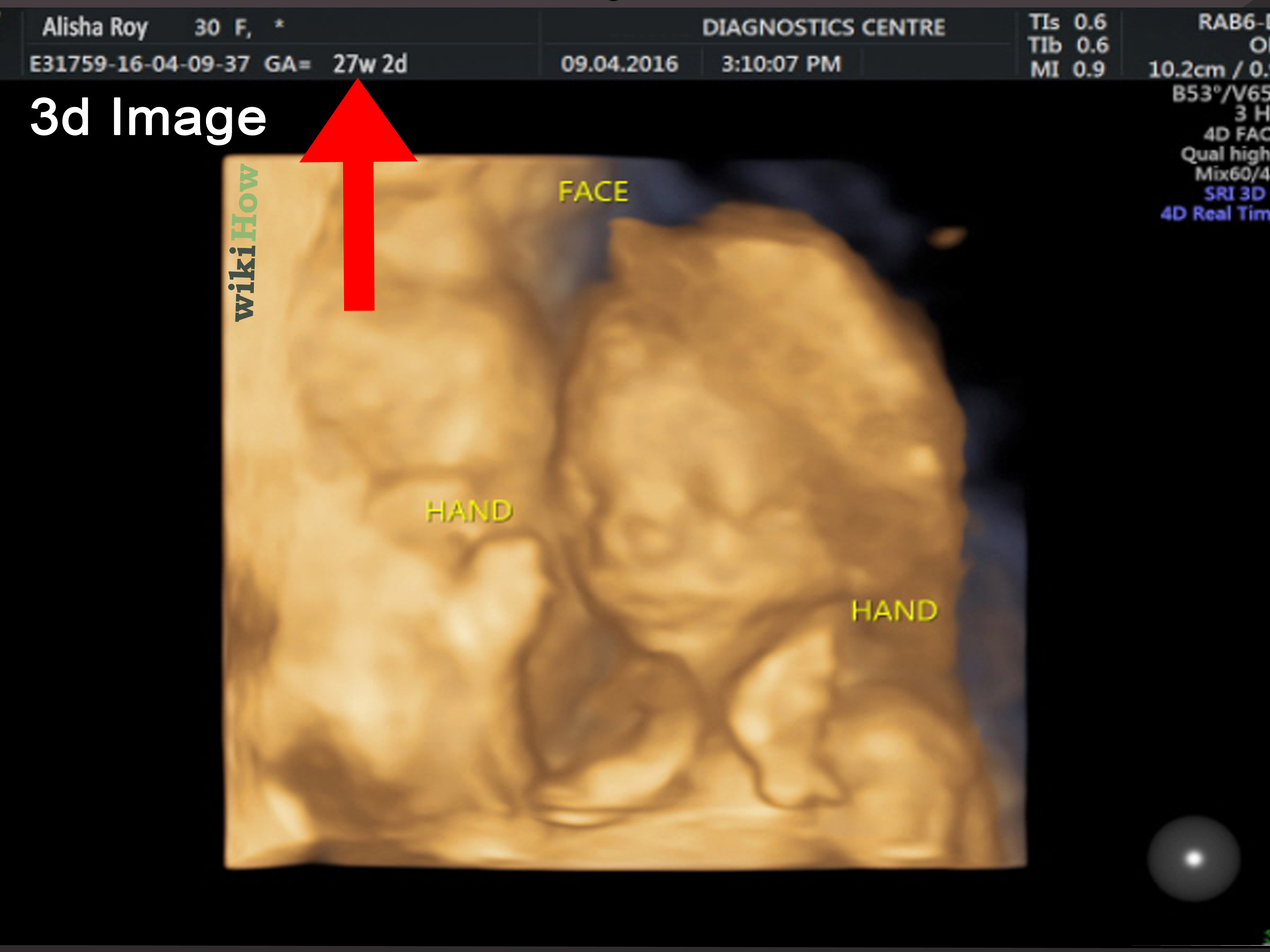
At a 20-week anatomy scan, the sonographer usually checks major structures such as the brain, spine, heart, stomach, kidneys, bladder, limbs, placenta, and umbilical cord. Parents may see a profile view, tiny feet, a spine that looks like a string of pearls, or a hand that appears to be giving a royal wave.
Abdominal Ultrasound
An abdominal ultrasound may evaluate the liver, gallbladder, pancreas, spleen, kidneys, aorta, and bile ducts. It can help investigate abdominal pain, swelling, abnormal blood tests, gallstones, kidney obstruction, liver changes, or aneurysm screening.
Fluid-filled structures often appear dark, while stones or calcifications may appear bright with shadowing. Organs have their own typical shapes and textures, which doctors compare with expected patterns.
Pelvic Ultrasound
A pelvic ultrasound may examine the uterus, ovaries, cervix, fallopian tube region, bladder, or nearby tissues. It may be performed externally over the lower abdomen or internally with a transvaginal probe, depending on the clinical need.
Pelvic ultrasound can help evaluate menstrual concerns, pelvic pain, ovarian cysts, fibroids, pregnancy-related questions, or abnormal bleeding. The images may show the uterus as a pear-shaped structure and ovaries with small follicles that may look like tiny dark circles.
Cardiac Ultrasound
An ultrasound of the heart is called an echocardiogram. It evaluates heart chambers, valves, wall motion, and blood flow. Doppler color is especially useful here because it helps show how blood moves through the heart.
To the untrained eye, an echocardiogram may look like a dramatic black-and-white movie starring a very busy jellyfish. To a cardiologist, it provides valuable information about heart structure and function.
Helpful Tips for Reading an Ultrasound Image More Confidently
Do Not Judge Image Quality Too Quickly
Ultrasound quality depends on many factors, including body position, bowel gas, fluid, tissue depth, fetal position, movement, and the type of probe used. A blurry image does not always mean a poor exam. Sometimes the body simply refuses to pose nicely.
Remember That Still Images Are Only Snapshots
During the actual exam, the sonographer views moving images in real time. A printed or digital still image may show only one selected moment. That is why the official report matters more than a single picture.
Use the Report as Your Main Guide
If you have access to both the ultrasound picture and the written report, use them together. The picture helps you visualize; the report explains what matters medically.
Ask Your Provider Before Searching Random Image Comparisons
Online image searches can create unnecessary panic because normal and abnormal findings may look similar to non-experts. Two ultrasound images can look alike but mean different things depending on context. Your doctor can explain what applies to your specific case.
Experience-Based Insights: What People Often Learn When Reading Ultrasound Pictures
Many people remember their first ultrasound as a mix of excitement, curiosity, and complete visual confusion. The screen lights up, the sonographer moves the probe, and suddenly everyone is staring at gray shapes with the intensity of detectives in a movie. Then the sonographer says, “There’s the head,” and half the room silently thinks, “Ah yes, of course,” while seeing what appears to be a potato in a snowstorm.
The first practical experience most people have is learning that ultrasound images are not photographs. They are sound-based slices of the body. That explains why a baby’s face might look perfectly clear in one frame and like a tiny ghost in the next. It also explains why organs may look unfamiliar even when they are completely normal. The machine is not showing skin, color, or realistic depth. It is showing how sound waves interact with tissue.
Another common lesson is that movement matters. During a pregnancy ultrasound, parents may see a heartbeat flicker, a hand move, or a baby turn away at the exact moment everyone wants a cute profile picture. During an abdominal ultrasound, the sonographer may ask the patient to hold their breath, roll slightly, or change position. These small movements help create better views. So if the exam feels like a gentle workout directed by someone holding a magic computer wand, that is normal.
People also learn that measurements are more meaningful than “what it looks like to me.” A round dark area might be normal fluid, a cyst, a follicle, a bladder, or something else entirely depending on location and context. A bright line might be bone, a tissue boundary, or an artifact. This is why experienced clinicians rely on multiple views, measurements, Doppler settings, and patient history instead of one dramatic screenshot.
Another helpful experience is comparing expectation with reality. Many patients expect ultrasound to provide instant answers. Sometimes it does. Other times, the result is “probably benign,” “needs follow-up,” or “limited view due to bowel gas.” That can feel frustrating, but it is part of responsible imaging. Medicine is careful because bodies are complicated, and ultrasound is excellent but not magical. It is more like a very smart flashlight than a crystal ball.
For expectant parents, one memorable lesson is that fetal position controls the show. A baby may hide their face, cross their legs, turn away, or wiggle constantly. This does not automatically mean a problem. It means the tiny star of the appointment has not read the schedule. Sonographers are trained to work around movement and position, but sometimes a follow-up scan is needed simply because the view was not complete.
People who have had abdominal, thyroid, breast, or pelvic ultrasound often learn the value of calm curiosity. It is natural to zoom in on every spot and wonder what it means. However, the better approach is to write down questions and discuss the official findings with a healthcare provider. Asking “What did the report say?” is usually more useful than asking “Why does this part look weird?” because ultrasound images can look strange even when they are normal.
Finally, many people discover that understanding the basics makes the experience less stressful. You do not need to become a radiologist to benefit from knowing that black often means fluid, white often means dense tissue, gray often means soft tissue, and color often shows motion or blood flow. With those basics, the ultrasound picture becomes less like alien weather and more like a medical map. You may not read every road sign, but you can at least tell where the major landmarks are.
Conclusion
Learning how to read an ultrasound picture can make medical imaging feel less mysterious and more empowering. Start with the labels, understand grayscale patterns, learn basic terms, check orientation, identify the main structure, review measurements, interpret Doppler colors carefully, compare the image with the written report, and remember the limits of self-interpretation.
An ultrasound picture can tell a fascinating story about the body, but the official interpretation belongs to trained professionals. Use your curiosity wisely, ask good questions, and let your healthcare team connect the dots. That way, you get the best of both worlds: informed confidence and expert guidance.